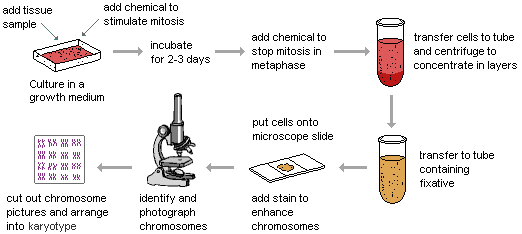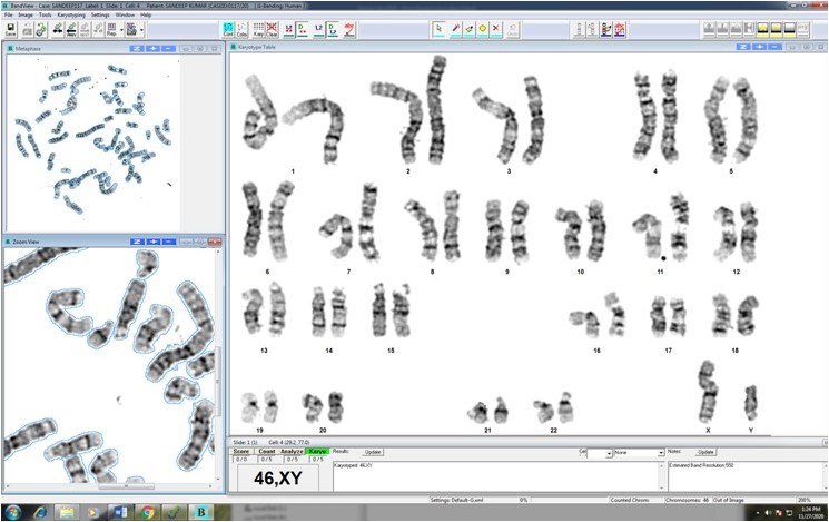
Duplicate cultures are initiated for each blood sample/ other sample types received for chromosome analysis (karyotype). Blood lymphocytes are cultured in complete media supplemented with the mitogen, phytohemeagglutinin (PHA) to divide and cultured for 72 hours routinely.Conventional Cell Culture Techniques followed by G-banding Chromosomes analysis through Bright Field Microscopy for detection of numerical and structural chromosome abnormalities including balanced and unbalanced translocations through GenASIs Bandview software from Applied Spectral Imaging (ASI).


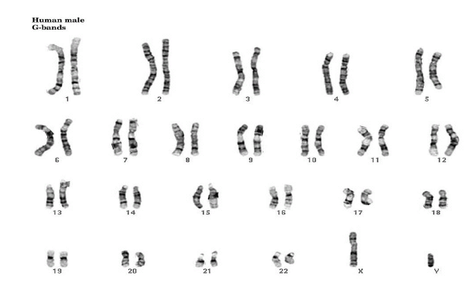
Figure 1: Chromosomes are stained and photographed for study and depicts 23 pairs or 46 chromosomes (each pair contributing 1 chromosome from father and 1 from mother) including pair of sex chromosomes with male showing one X chromosome and one Y chromosome (Normal Male Karyotype: 46, XY.

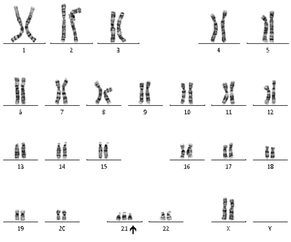
Figure 2: Abnormal Karyotype of child showing Trisomy of chromosomes #21 (Downs Syndrome) due to extra copy of chromosome 21. (Abnormal Female Karyotype: 47, XX, +21).

Methodology:Conventional Cell Culture Techniques followed by G-banding Chromosomes analysis through Bright Field Microscopy for detection of numerical and structural chromosome abnormalities including balanced and unbalanced translocations through GenASIs Bandview software from Applied Spectral Imaging (ASI).